
Services
Our Services
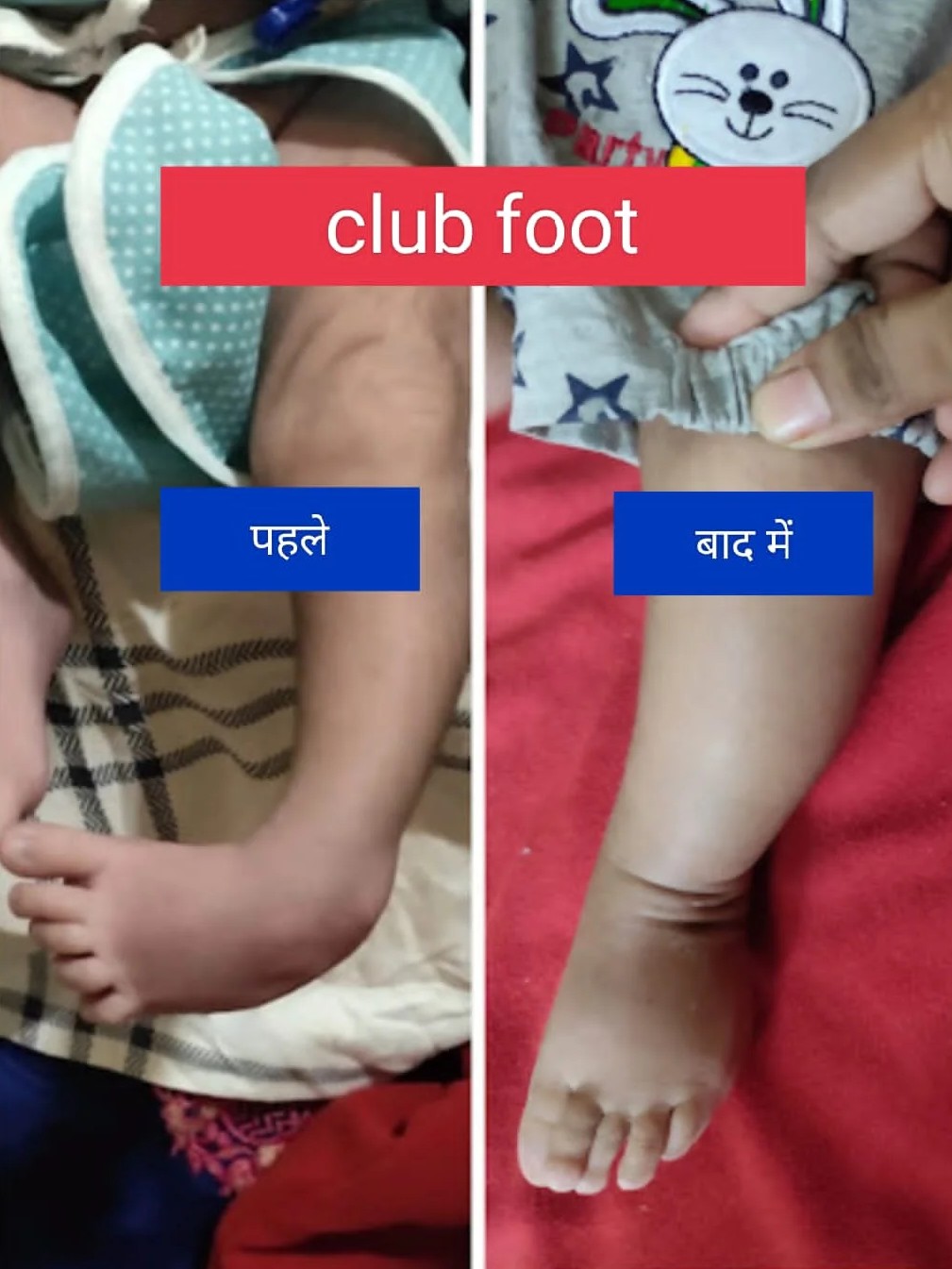
Club Foot
Clubfoot, also known as talipes equinovarus, is a congenital deformity of the foot that affects approximately 1 in 1,000 births.
Causes and Risk Factors
1. Genetic predisposition
2. Environmental factors (e.g., intrauterine positioning)
3. Associated conditions (e.g., cerebral palsy, spina bifida)
4. Family history
Characteristics
1. Inward rotation of the heel (equinus)
2. Upward rotation of the foot (dorsiflexion)
3. Inward rotation of the ankle (varus)
4. Adduction of the forefoot (toe-in)
5. Rigidity and stiffness
Classification
1. Idiopathic (most common, 80-90%)
2. Neurogenic (associated with neurological conditions)
3. Syndromic (part of a larger genetic syndrome)
Treatment Options
Non-Surgical
1. Ponseti Method: Serial casting and manipulation (90-95% success rate)
2. Kite Method: Similar to Ponseti, with modifications
3. Physical therapy and bracing
Surgical
1. Posteromedial release surgery (PMRS): Release of tight tissues and tendons
2. Osteotomies: Cutting and realigning bones
3. Tendon transfers
Ponseti Method
1. Initial casting (1-2 weeks)
2. Weekly casting changes (4-6 weeks)
3. Achilles tendon tenotomy (if necessary)
4. Bracing (2-3 years)
Surgical Indications
1. Failed non-surgical treatment
2. Severe deformity
3. Older children or adults
4. Associated conditions (e.g., cerebral palsy)
Complications
1. Recurrence
2. Infection
3. Nerve damage
4. Limited mobility
Outcomes
1. Successful correction in 90-95% of cases
2. Improved mobility and function
3. Enhanced quality of life
4. Potential for long-term follow-up and management
Radial Club Hand
Radial club hand, also known as radial deficiency, is a rare congenital condition that affects the development of the radius bone in the forearm.
This results in a shortened or missing radius, leading to a curved or “club-like” appearance of the hand.
characteristics - – Shortened or missing radius bone – Curved or bent wrist and forearm – Shortened or missing thumb – Fusion or webbing of fingers – Limited wrist and hand movement
Treatment options-
1. Serial plaster to correct deformity –
2 Surgery to straighten the wrist and improve hand function –
3-Physical therapy to improve mobility and strength –
4-Orthotics or prosthetics to support hand function –
5-Occupational therapy to develop adaptive skills
Foot Deformities
Foot deformities are conditions where the foot has abnormal shape, position, or alignment, affecting movement and walking.
Flat foot
Flat foot is a condition where the arch of the foot collapses, causing the sole to touch the ground while walking daily.
Vertical Talus
Congenital Vertical Talus (CVT), also known as Rocker Bottom Foot or Congenital Vertical Talus Deformity, is a rare congenital foot anomaly. Incidence and Prevalence 1. Estimated 1 in 10,000 to 1 in 50,000 births
2. More common in males (2:1 ratio)
3. Bilateral involvement in 50-60% of cases
Causes and Risk Factors
1. Genetic predisposition
2. Chromosomal abnormalities (e.g., trisomy 13, 18)
3. Neural tube defects (e.g., spina bifida)
4. Intrauterine constraints or positioning
5. Family history
Characteristics
1. Vertical orientation of the talus bone
2. Dorsal dislocation of the navicular bone
3. Plantarflexion of the forefoot
4. Hindfoot equinus
5. Rigid or flexible deformity Classification
1.-Idiopathic (most common)
2. Neurogenic (associated with neurological conditions) 3. Syndromic (part of a larger genetic syndrome)
Diagnosis
1. Physical examination
2. Imaging studies: – X-rays ( AP and lateral views) – CT scans or MRI (for complex cases)
3. Differential diagnosis: other congenital foot deformities (e.g., clubfoot) Treatment Options
Non-Surgical 1. Manipulation and casting (Ponseti method)
2. Physical therapy and bracing 3. Orthotics (e.g., ankle-foot orthoses)
Surgical
1. One-stage correction (dorsal opening wedge osteotomy)
2. Two-stage correction (initial casting, followed by surgery)
3. Osteotomies (e.g., calcaneal lengthening)
4. Tendon transfers or lengthening Complications
1. Recurrence
2. Infection
3. Nerve damage
4. Limited mobility
5.Arthritis or degenerative changes
Outcomes
1. Successful correction in 80-90% of cases
2. Improved mobility and function
3. Enhanced quality of life
4. Potential for long-term follow-up and management
Torticollis
Torticollis, also known as wry neck, is a condition characterized by a twisted or tilted neck, resulting in an abnormal postural position. Overview Types: 1. Congenital Muscular Torticollis (CMT): Present at birth, caused by in-utero positioning or genetic factors.
2. Acquired Torticollis: Develops later in life due to various factors.
3. Spasmodic Torticollis: A neurological disorder causing involuntary muscle contractions.
Causes and Risk Factors:
1.Congenital factors (e.g., intrauterine positioning)
2. Genetic predisposition
3.Trauma or injury
4. Infections (e.g., mastoiditis)
5. Neurological conditions (e.g., cerebral palsy)
6. Musculoskeletal conditions (e.g., muscle imbalances) Symptoms: 1. Twisted or tilted neck
2. Limited range of motion
3. Head tilt or rotation
4. Facial asymmetry
5. Pain or stiffness
6. Difficulty turning head
Diagnosis:
1. Physical examination
2. Imaging studies (e.g., X-rays, CT scans, MRI)
3. Electromyography (EMG) for spasmodic torticollis
Treatment : Conservative: 1. Physical therapy (stretching, strengthening)
2. Occupational therapy (positioning, activities) 3. Orthotics (e.g., collars, splints) 4. Pain management (medications, injections)
Surgical: 1. Muscle lengthening or release 2. Tendon transfers
3. Osteotomies (bone realignment)
4. Spinal fusion (in severe cases)
Complications: 1. Permanent deformity
2. Chronic pain
3. Limited mobility
4. Facial asymmetry
5. Psychological impacts (e.g., self-esteem)
Polydactyly
Polydactyly is a birth condition where a person has extra fingers or toes, which may be fully formed or small and fused.
Aneurysmal bone cysts
Aneurysmal bone cysts are benign, blood-filled bone lesions that cause swelling, pain, and weaken the affected bone.
Radioulnar Synostosis
Radioulnar synostosis is a birth condition where the radius and ulna are fused, limiting forearm rotation movement.
Humerus Varus
Humerus varus is a deformity where the upper arm bone angles inward, reducing shoulder alignment and arm movement.
Knock Knee
Knock knee, also known as genu valgus, is a condition where the knees touch or nearly touch each other, causing a gap between the ankles.
It’s common in children and often corrects itself with growth.
Causes:
1. Genetic predisposition
2. Growth and development
3. Injury or trauma
4. Infections (e.g., osteomyelitis)
5. Neuromuscular disorders (e.g., cerebral palsy) Symptoms:
1. Knees touch or nearly touch
2. Gap between ankles
3. Uneven gait or walking pattern
4. Difficulty standing or balancing
5. Pain or discomfort in knees, legs, or feet
Treatment:
1. Observation and monitoring
2. Bracing or orthotic
3. Physical therapy
4. Surgery (in severe cases)
Exercises to help correct knock knee:
1. Strengthening exercises (leg lifts, squats, lunges)
2. Stretching exercises (hamstring, quadriceps)
3. Balance and coordination exercises
When to seek medical attention:
1. Severe knock knee
2. Pain or discomfort
3. Difficulty walking
4. Uneven growth or development
5. Concerns about appearance or self-esteem
Knee Deformities
Flexion deformity--It is called when knee is bend and cannot be straighten
straightened knee are required for standing and normal walking
Treatment--inital stgaes
Exercises
Moderate deformity--plaster cast
Severe Deformity --Surgery
Cerebral palsy
Orthopedic surgery is commonly performed in cerebral palsy (CP) patients to address musculoskeletal issues that affect mobility, posture, and overall quality of life.
Common Orthopedic Issues in CP Patients
1. Muscle spasticity and contractures
2. Hip subluxation or dislocation
3. Knee flexion or extension contractures
4. Foot deformities (e.g., equinus, varus, or valgus)
5. Scoliosis and other spinal deformities
6. Osteoporosis and fractures
Surgical Goals
1.Improve mobility and function
2. Enhance posture and balance
3. Reduce pain and discomfort
4. Prevent or correct deformities
5. Improve overall quality of life Common Orthopedic Surgeries
1. Muscle lengthening procedures– Lengthening of muscles and tendons to reduce spasticity and contractures.
2. Tendon transfers– Transferring tendons to improve muscle balance and function
. 3. Osteotomies– Cutting and realigning bones to correct deformities.
4. Hip reconstruction Surgery to correct hip subluxation or dislocation.
5. Spinal fusion Surgery to correct scoliosis or other spinal deformities.
6. Foot correction Surgery—to correct foot deformities.
7. Bone stabilization Surgery to stabilize bones and prevent fractures.
Considerations and Challenges
1. Complexity of CP- Each patient’s condition is unique, requiring individualized treatment plans.
2.Multiple surgeries- CP patients may require multiple surgeries throughout their lifetime.
3.Anesthesia risks- Patients with CP may have increased anesthesia risks due to associated medical conditions.
4. Postoperative care: Requires specialized rehabilitation and therapy to optimize outcomes.
5. Growth considerations– Surgeons must consider growth plates and potential impact on future growth.
Preoperative Evaluation
1. Comprehensive medical history and physical examination
2. Imaging studies (e.g., X-rays, CT scans, MRI)
3.Assessment of cognitive and communication abilities
4. Evaluation of nutritional status and bone health
5. Consultation with rehabilitation team and other specialists
Postoperative Rehabilitation
1. Pain management
2. Physical therapy to maintain range of motion and strength
3. Occupational therapy to improve daily functioning
4. Orthotics and assistive devices to support mobility and balance
5. Regular follow-up with orthopedic surgeon and rehabilitation team Outcomes and Research
1. Improved mobility and function
2. Reduced pain and discomfort
3. Enhanced quality of life
4. Prevention of deformities and complications
5. Ongoing research focuses on optimizing surgical techniques, improving outcomes, and reducing complications.
Your Child’s Care and Comfort Come First.
If Your child is suffering from any kind of Bones & Joint problem
Dr Vivek Shrivastava offers child-friendly pediatric orthopedic care.
Reach Us
© 2025 Dr. Vivek Shrivastava. All Rights Reserved.


